Week of 1 December 2020

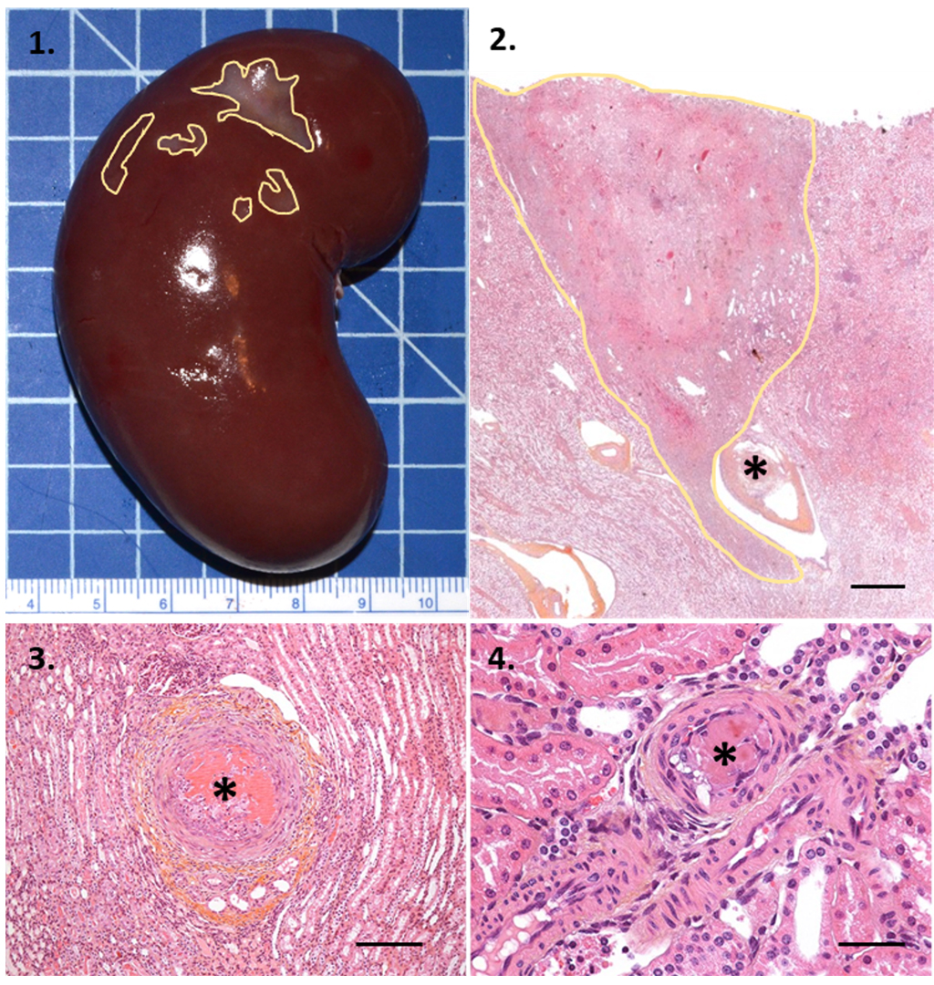
Multiple infarcts (areas outlined in yellow) occurred in this kidney!
Renal infarcts are generally due to a thromboembolic event, a possible complication of cardiovascular surgery or interventional procedures performed on the left side of the heart or supra-renal aorta. It is rarely possible to observe the thrombus (*) that caused the infarct in the histologic sections.
1. Macroscopic view at explantation.
2. HE&S, original magnification: x1, scale bar: 1 mm.
3. HE&S, original magnification: x10, scale bar: 250 µm.
4. HE&S, original magnification: x20, scale bar: 125 µm.
Yellow line: Delimitation of renal infarcts.
*Thrombus.
This histopathology image is one example from the comprehensive suite of Pathology Services offered by IMMR’s in-house team of Board-certified Veterinary Pathologists.
Contact us to learn more and discuss your preclinical research and pathology evaluation needs.
Follow us on LinkedIn and don’t miss new images from our library that we post every Tuesday, when you’ll have another chance to recognize, identify or diagnose what is shown. You can also stay updated on some of the latest developments in Preclinical Science. Stay tuned!


