Week of 24 August 2021

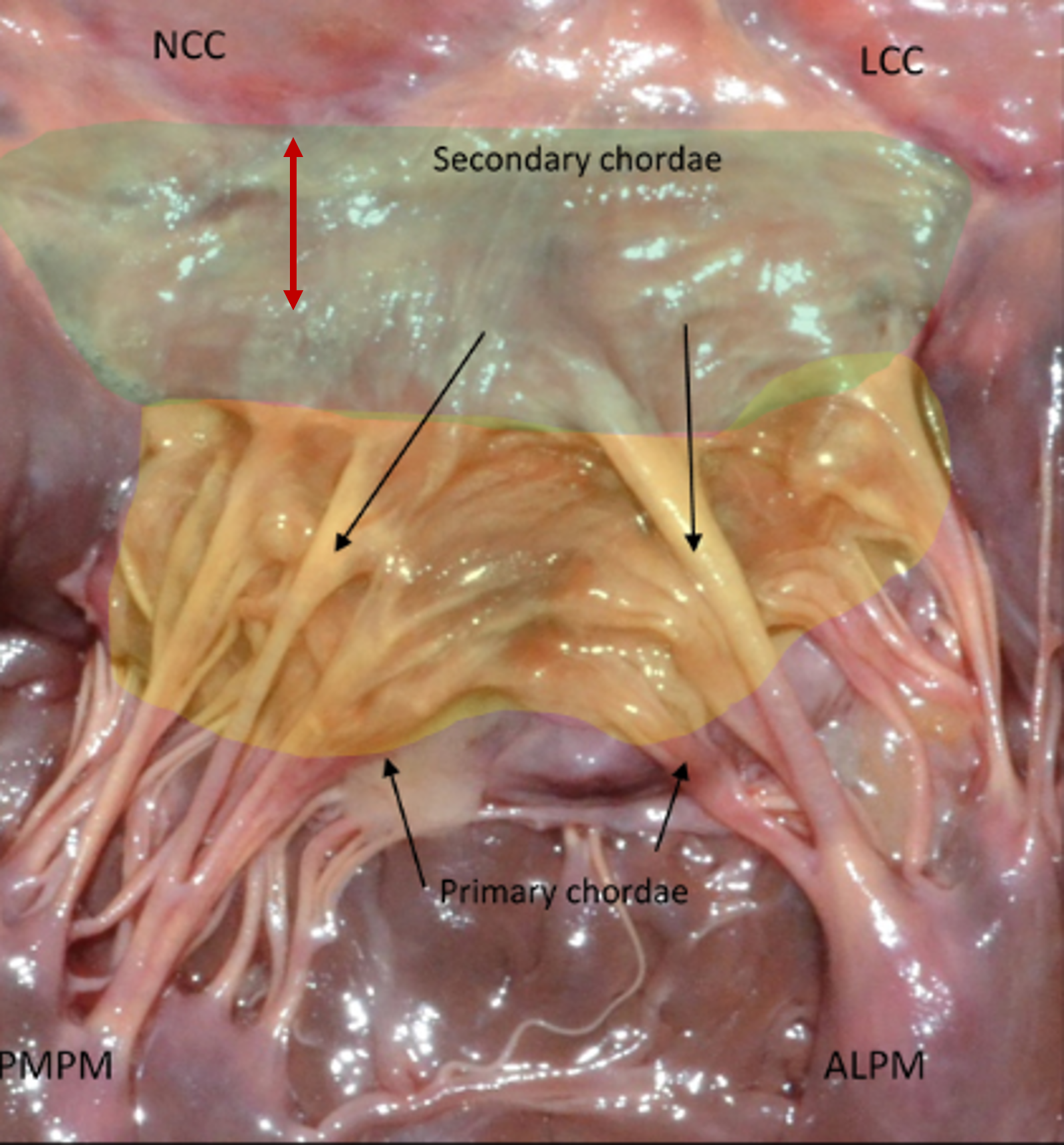
This image is a photograph of a mitral valve of a human!
A major difference among the species we have seen in this ‘Comparative anatomy of the mitral valve’ series, is the presence of a true aorto-mitral “curtain” along the human anterior leaflet, below the mitral annulus, defined as a space between the mitral and aortic valves.1
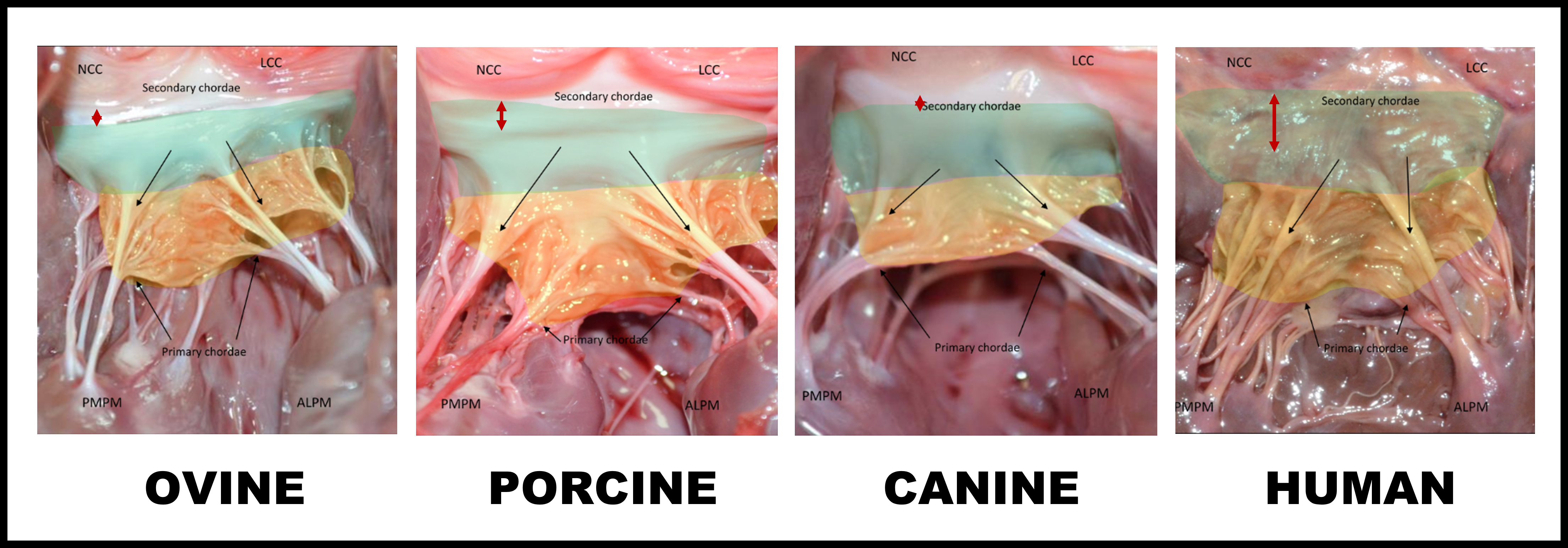
The colors are added to indicate the rough zone of the anterior leaflet (yellow) and the smooth zone of the anterior leaflet (green).
NCC: Noncoronary cusp.
LCC: Left coronary cusp.
ALPM: Anterolateral papillary muscle.
PMPM: Posteromedial papillary muscle.
Red double headed arrow: Aorto-mitral “curtain”.
You can now compare the images of the previous weeks to each other:

As shown here, and as you noticed through the past four weeks, there are important anatomical differences between sheep, pigs, and dogs, explaining some of the challenges associated with transcatheter mitral procedures in large animal models. Comparative anatomy is key to understanding which models are best, their potential limitations and how to leverage this information to provide compelling evidence of safety and efficacy of a novel device.
These photographs are examples from the comprehensive suite of Pathology Services offered by IMMR’s in-house team of Board-certified Veterinary Pathologists.
Contact us to learn more and discuss your preclinical research and pathology needs.
Follow us on LinkedIn and don’t miss new images from our library that we post every Tuesday, when you’ll have another chance to recognize, identify or diagnose what is shown. You can also stay updated on some of the latest developments in Preclinical Science. Stay tuned!
1 Borenstein N, Behr L, Morlet A, Chevènement O, Kieval R, Ente A, Fiette L: Large animal models for the interventional cardiologist: a comparative anatomy, imaging, histopathology and regulatory perspective. The PCR-EAPCI Textbook, Part IV.


